Home

Moschee Preşedinte evaluabile sarcomere electron microscopy Nouălea deasupra a lega

Transmission electron microscopy image of the sarcomere, with... | Download  Scientific Diagram
Transmission electron microscopy image of the sarcomere, with... | Download Scientific Diagram

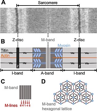
Myomesin - Wikipedia

330 Electron Microscopy and the The Sliding Filament Model of Skeletal  Muscle Contraction - YouTube
330 Electron Microscopy and the The Sliding Filament Model of Skeletal Muscle Contraction - YouTube

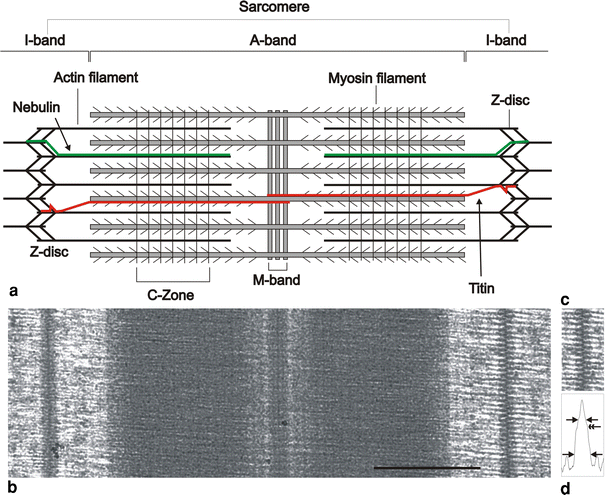
a) Electron micrograph showing a whole sarcomere from fish muscle in... |  Download Scientific Diagram
a) Electron micrograph showing a whole sarcomere from fish muscle in... | Download Scientific Diagram

Sarcomere location of MyBP-C. The electron micrograph was taken from... |  Download Scientific Diagram
Sarcomere location of MyBP-C. The electron micrograph was taken from... | Download Scientific Diagram

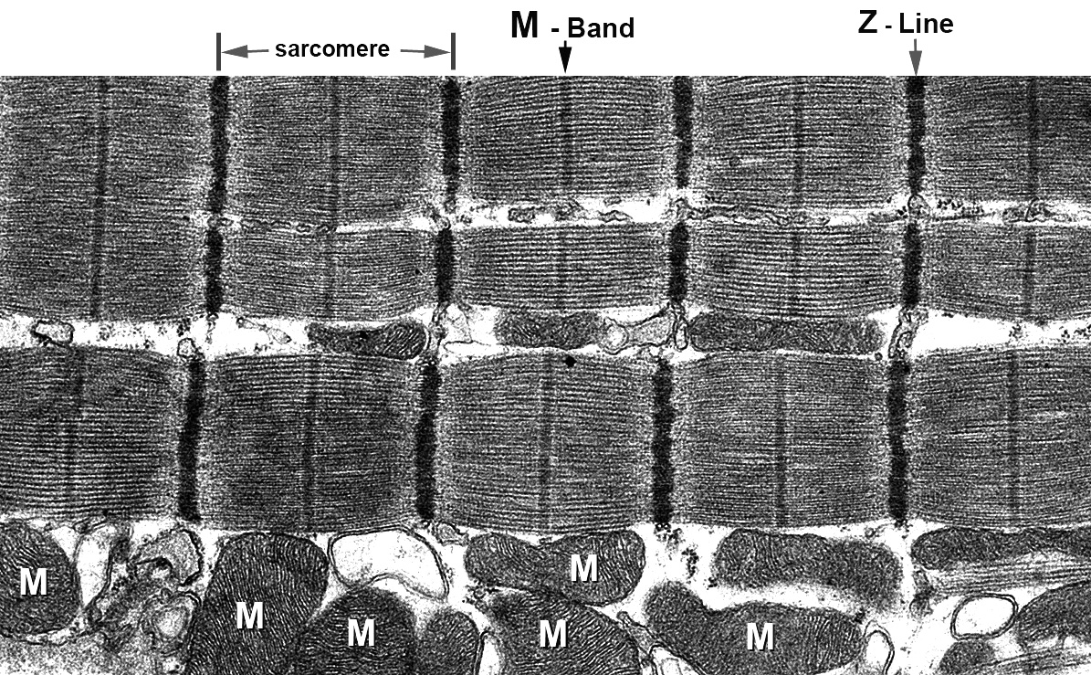
Transmission electron micrograph of a sarcomere. Transmission electron... |  Download Scientific Diagram
Transmission electron micrograph of a sarcomere. Transmission electron... | Download Scientific Diagram

B1 Muscles and Movement | Brent Cornell

330 Electron Microscopy: the Sliding Filament Model of Skeletal Muscle  Contraction - YouTube
330 Electron Microscopy: the Sliding Filament Model of Skeletal Muscle Contraction - YouTube

PDF] Muscle Contraction. | Semantic Scholar

Histology Laboratory Manual

Muscle: The Histology Guide

Losing pieces without disintegrating: Contractile protein loss during  muscle atrophy | PNAS
Losing pieces without disintegrating: Contractile protein loss during muscle atrophy | PNAS

Polarization-resolved microscopy reveals a muscle myosin motor-independent  mechanism of molecular actin ordering during sarcomere maturation | PLOS  Biology
Polarization-resolved microscopy reveals a muscle myosin motor-independent mechanism of molecular actin ordering during sarcomere maturation | PLOS Biology
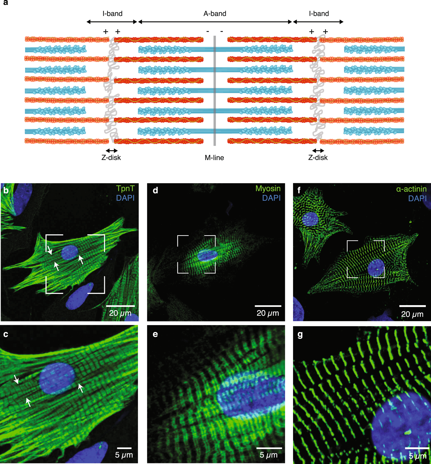
The vertebrate muscle Z-disc: sarcomere anchor for structure and signalling  | SpringerLink
The vertebrate muscle Z-disc: sarcomere anchor for structure and signalling | SpringerLink

Muscle tissue - Knowledge @ AMBOSS

Molecular plasticity of the native mouse skeletal sarcomere revealed by  cryo-ET | bioRxiv
Molecular plasticity of the native mouse skeletal sarcomere revealed by cryo-ET | bioRxiv

Molecular-scale visualization of sarcomere contraction within native  cardiomyocytes | Nature Communications
Molecular-scale visualization of sarcomere contraction within native cardiomyocytes | Nature Communications

Cancer Histology Core

330 Electron Microscopy and the The Sliding Filament Model of Skeletal  Muscle Contraction - YouTube
330 Electron Microscopy and the The Sliding Filament Model of Skeletal Muscle Contraction - YouTube

Polarization-resolved microscopy reveals a muscle myosin motor-independent  mechanism of molecular actin ordering during sarcomere maturation | PLOS  Biology
Polarization-resolved microscopy reveals a muscle myosin motor-independent mechanism of molecular actin ordering during sarcomere maturation | PLOS Biology
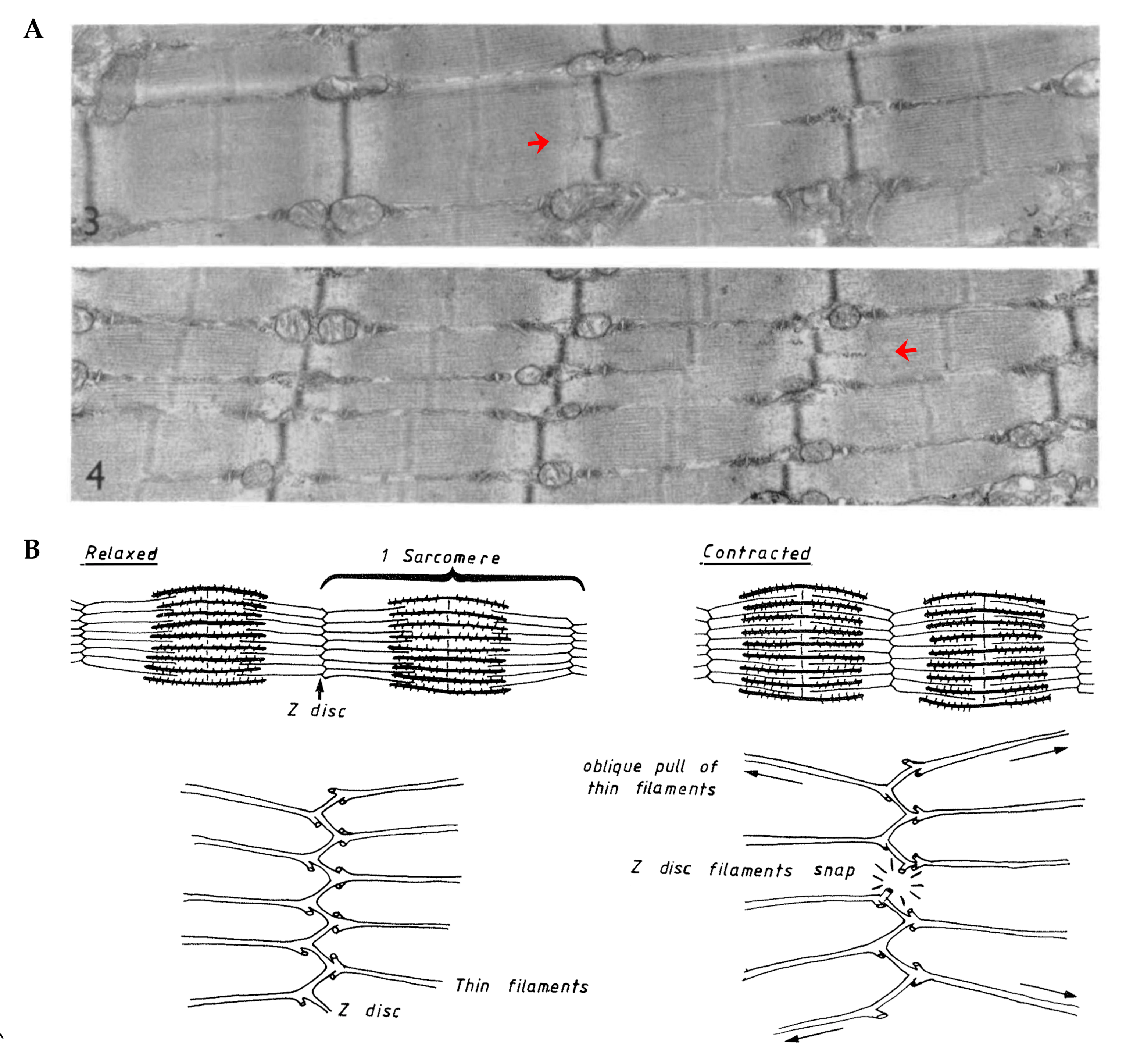
4 Electron microscopy picture of the sarcomere. | Download Scientific  Diagram
4 Electron microscopy picture of the sarcomere. | Download Scientific Diagram

The Titin/Telethonin Complex

Cells | Free Full-Text | Identifying the Structural Adaptations that Drive  the Mechanical Load-Induced Growth of Skeletal Muscle: A Scoping Review
Cells | Free Full-Text | Identifying the Structural Adaptations that Drive the Mechanical Load-Induced Growth of Skeletal Muscle: A Scoping Review

Figure 5 from The organization of titin filaments in the half-sarcomere  revealed by monoclonal antibodies in immunoelectron microscopy: a map of  ten nonrepetitive epitopes starting at the Z line extends close to
Figure 5 from The organization of titin filaments in the half-sarcomere revealed by monoclonal antibodies in immunoelectron microscopy: a map of ten nonrepetitive epitopes starting at the Z line extends close to